As many of you know, I'm working on an ORCA research project and my honors thesis. Both projects involve making a microfluidic device and preterm birth. One of them is trying to figure out why a certain protein tends to be in higher concentrations in a woman's blood when she is going to deliver preterm. The other is making a device that a doctor could use to predict whether or not a pregnancy was going to end in premature labor. (Not including trauma induced premature labor.)
Here are a few pictures from around the lab(s):
 |
| After I print the designs onto the templates for my devices in the clean room (in the Clyde building) I etch it in this hood so the design can be embossed into the device. |
 |
| Several molds are made out of a single wafer, so we break them apart and inspect the templates here. |
 |
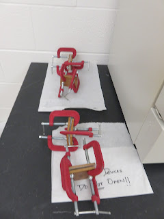
| You can see the molds in the petri dish at the top of this photo. The green lines are what's going to be imprinted into the device to make the microfluidic channels. The square rectangle to the left of the razor blade in the center is a paper covered piece of PMMA, a substance similar to glass and plastic. I use the razor blade to take the paper off, wash it well in IPA and distilled water and then compress it with a template between 2 glass slides and 2 copper plates (pictured to the right of the center razor blade). The contraption is then held together with 4 C-clamps. |
 |
| This is where I dry my PMMA off and put them in their glass slide/copper plate sandwiches. |
 |
I use this drill to drill reservoirs into the cover plate. The cover plates are then put side by side into a glass slide/copper plate sandwich held together by C-clamps as well.
 |
| This is what they look like when they've had their holes drilled and the edges cleaned up. |
 |
Once they're clamped together, a cover plate/glass slide/copper plate sandwich and 3 template/plastic/glass slide/copper plate sandwiches go in this oven at 138 degrees for 25 minutes or so to imprint the channels into the slides and flatten the cover plates.

Once they cool, I align a cover plate that's had holes drilled into it and a glass slide that's had the channels imprinted into it, sandwich them between glass slides and copper plates, clamp it all together with C-clamps, and put them in this oven for another 25 minutes or so at 110 degrees to bond the two pieces together.
 |
This is what they look like when they come out of the oven and are cooling.
This is what the finished devices look like. (Sorry, it's an old pic of a well used device from my cell phone)
|
 |
| This white microscope is the microscope I use to inspect my devices and make sure the channels aren't compromised. |
 |
This is the lab bench where I test the channels with IPA and clean them on vacuum suction overnight. The smallest particle can clog a device so gloves are always warn and the utmost care is taken to keep them clean.
 |
| I use this bench to make buffer solutions and to label proteins and peptides. |
 |
| This is part of our chemical supply. (Not pictured includes: samples that have to be refrigerated and flammables) |
 |
| This is where I store my labeled samples and buffers between use. |
|
 |
| This is the bench top and microscope I use to run my separation e |
 |
| This is a close up of the microscope/laser combination I use to electrophoretically separate my PTB biomarkers and quantify them by detecting the light produced when a laser hits the florescent tags I put on my proteins and peptides. |


No comments:
Post a Comment